
INTRODUCTION
At present, patients are more interested in dental treatments with better esthetic results and less treat-ment time. Rehabilitation of occlusion with dental im-plants is considered one of the most efficient treatment methods for edentulism.1 While implant dentistry has become a desirable option for replacement of missing teeth, the available bone foundation determines the possibility of implantation.2
Lack of sufficient bone to place an implant at the functionally and aesthetically most appropriate position is a common problem.3, 4 To facilitate osseointegration and avoid bone resorption, narrow, edentulous alveolar bony ridges less than 5-mm wide require bone augmentation prior to implant placement.2 By augmenting the defective area, the implant surgeon provides the necessary foundation for implant placement by establishing a bony wall thickness of at least 1 mm around screw-type implants.5, 6 Traditional methods of ridge expansion include onlay and inlay bone grafts, sandwich osteotomies, guided bone regeneration, and alveolar distraction osteogenesis.6
Crestal ridge bone augmentation is an alternative bone expansion technique that can be used to augment the atrophic maxilla and mandible prior to implant placement. This method was first introduced by Dr Hilt Tatum in the 1970s and was commonly referred to as ridge splitting, bone spreading, or ridge expansion technique.7 This method aims at the generation of bone around the implant sites by bone osteotomies that enable buccal cortex repositioning after greenstick fracture of the buccal bony wall. Since its introduction, many studies have attempted to prove that the alveolar ridge split technique is a good alternative to traditional alveolar augmentation procedures.8-10
Treatment of atrophic ridge especially in posterior man-dibular is accompanied with great problem in achiev-ing successful results with endosseous implants.3, 4
According to Atwood knife-edge crests might be managed by conventional bone grafting, guided bone augmentation procedures by using membranes, and various other techniques.11 Ridge augmentation by bone graft requires a second surgery for a later implantation, thus lengthening the treatment time and cost. Ridge splitting technique which causes lateral ridge expansion creates new im-plant bed by longitudinal osteotomy positioning buc-cal cortex laterally.5
The present clinical report describes the technique for ridge splitting, gradual expansion in the mandible and simultaneous implant placement within the split ridge.
CASE REPORT
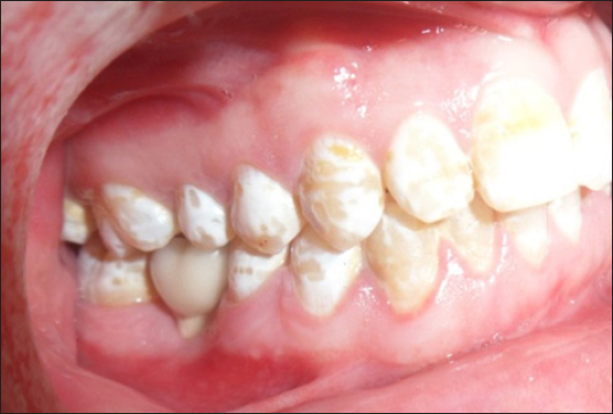
A 24-year-old systemically healthy male patient reported to the Department of Periodontology, Kamineni Institute of Dental Sciences, Narketpally, Telangana, with a chief complaint of bleeding gums and missing tooth number of 46. Patient’s medical history was noncontributory. Extra-and intraoral examinations had normal findings, and his dentition was in a good state of repair. Dental history revealed missing mandibular right teeth number 46, which had been extracted five years ago. Radiographic and clinical examination revealed inadequate buccolingual dimension of bone at the crest for implant placement, with sufficient height (Figure 1). There was adequate cortical and cancellous bone to allow ridge expansion. It was decided to place immediate implants, using the split control expanding technique. After administrating adequate local an-esthesia, a mid-crestal incision and intracrevicular incisions were made around buccal aspect of adjacent teeth. Full thickness mucoperi- osteal flaps were raised on the buccal and lingual aspects of cortical plates but minimal tissue reflection was per-formed in lingual aspect to preserve the periosteum at-tachment surrounding the buccal and lingual bone (Figure 2). This was performed to prevent possible buccal bone plate crack. Keeping the periosteum intact would facilitate re-positioning of the fragments and achieve good healing. Corticotomy was performed with a pizeotome facilitating the os-teotomy of region. The horizontal osteotomy line was cut along the narrow crest using a piezotome burs, 1 to 2 mm away from the second molar till the first premolar region on the right side of the mandible under saline irrigation. Then the osteotomy line was deepened. Two additional vertical cuts were created on the buccal plate at the mesial and distal end of the horizontal incision. Osteotomes of increasing size and chisels were used for the progressive lateralization of the buccal plate. A sequence of ex-pansion drill of increasing width in the selected site was used to allow more gradual bone expansion. Then the implant site was prepared using final twist drills and implant of 4.2 mm x 10 mm was placed in molar region. Allograft material was used to fill the space and resorbable membrane is placed and it is stabilized by cover screw (Figure 3). Then tension free mucoperiosteal tis-sue closure was performed over implant using 3-0 non--resorbable suture (Figure 4) Non steroidal analgesics, Amoxicillin 500 mg and 0.2% chlorhexidine mouth rinse was the pre-operative protocol administered for the patient. Sutures were removed after 10 days. After 3months prosthesis is placed (Figure 5)
|
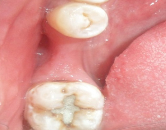
Figure 1: Clinical picture showing inadequate buccolingual dimension of bone at the crest for implant placement
Click here to view |
|
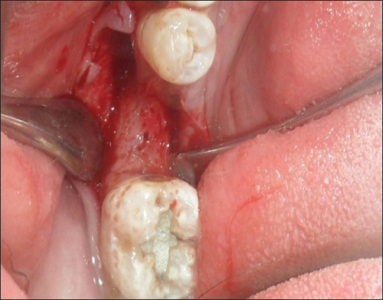
Figure 2: Operative picture showing after flap elevation
Click here to view |
|
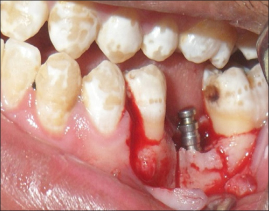
Figure 3: Allograft material was used to fill the space and resorbable membrane is placed and it is stabilized by cover screw
Click here to view |
|
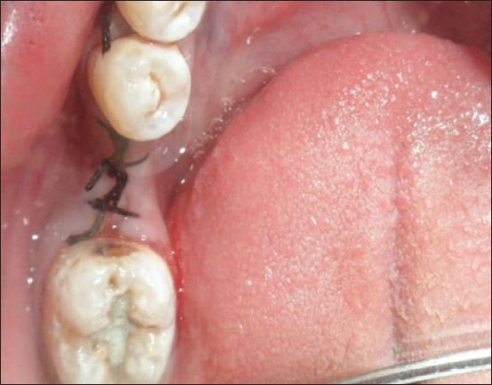
Figure 4: Tension free mucoperiosteal tissue closure was performed over implant using 3-0 non--resorbable suture
Click here to view |
|
Figure 5: Sutures were removed after 10 days and after 3months prosthesis is placed.performed with immediate implant placement,
Click here to view |
A significant increase was achieved in the bone dimen-sion, which enabled the placement of endosseous dental implant successfully. Then after preparation of the im-plant site, implant of 4.2 mm x 10 mm, was placed in molar region. The present report demonstrated the successful use of expanding the poste-rior mandibular alveolar ridge. It also showed that this technique allows for immediate implant placement.
DISCUSSION
Vari-ous surgical widening techniques have been described, including lateral augmentation with or without guided bone regeneration (GBR), ridge expansion osteotomy, ridge splitting technique with or without interpositional grafting and horizontal distraction osteogenesis.12-19
The buccal cortex is positioned laterally to create space between buccal and lingual cor-tical plates, which is filled by endosseous implant with or without any graft material.6, 20 This technique is which decreases the treatment time significantly.
A staged approach to ridge splitting in the mandible can be performed to avoid complications. Another tech-nique for placement of dental implants in narrow bone ridges is repositioning and remodeling of alveolar bone by controlled expansion. This technique uses screw-type configuration osteotomes and thread formers with increasing diameters.21
Horizontal atrophy of the alveolar ridge usually complicates adequate implant placement. GBR, bone grafting, alveolar ridge splitting and combinations of these techniques have been suggested as treatment modalities to increase bucco-lingual dimension of the residual ridge.7, 9, 21-31 The ridge splitting technique is used for the horizontal augmentation of narrow alveolar ridges and allows simultaneous implant placement. Low morbidity and short treatment time are the major advantages of this technique compared to GBR and bone grafting procedures.5, 32-34
The posterior mandible is the most difficult region for reconstruction and early implantation in cases of severe alveolar resorption in the maxilla- mandibular complex. Onlay grafting with biodegradable membranes and au-tografts is the most frequently used technique; however, this technique involves a long ossification period, and the tendency of the graft material to resorb can easily decrease bone quality and quantity.35 Time lost and donor-side morbidity are the main disadvantages of this reconstructive approach. The split-crest technique should be delineated as a bone expansion procedure, which potentially eliminates the overall disadvantages of Onlay grafting for esthetic and functional demands.36 Chiapasco et al. evaluated the efficiency of different sur-gical techniques for ridge reconstruction and success rates of implants placed in the augmented areas.11
The surgical success and the implant survival rates were as high as the guided bone regeneration and onlay graft procedure, with the advantage of a shorter treatment time.11 Careful preparation of the bone and maintenance of an attached periosteum are critical to the formation of new bone around the interproximal surfaces of the implants. Wound healing in these cases is similar to the fracture repair of bone. The gap is filled with a blood clot, which is organized and replaced with woven bone and further matures into load-bearing lamellar bone at the implant interface.35
Strong evidence for the effectiveness and the predictability of the ridge splitting technique is available in the literature. Clinical trials have reported success rates ranging from 98 to 100%. The survival rates of implants immediately placed in expanded sites ranged from 91% to 97.3%, while the success rates varied from 86.2% to 98.8% .8,9,16,37,38
Complications during the surgical procedure are very rare - fracture of the buccal bone plate being reported as a major complication of the technique.39 Controlled force application and gradual expansion could prevent malfractures.40, 41 In addition, a thorough preoperative evaluation is very important. The thickness of the cortical plates and the amount of the intervening cancellous bone must be carefully assessed preoperatively by dental CT scans. Last but not least, fabrication of radiographic/ surgical guide can prevent improper implant placement and angulation. If favorable condi-tions are not present, clinician might prefer Onlay aug-mentation. Therefore, appropriate case selection and sur-gical technique is of great importance when considering the application of this technique.
CONCLUSION
The ridge splitting technique seems to be a minimally invasive option for horizontal augmentation of narrow alveolar ridges. Predictable clinical results can be achieved as long as a proper preoperative evaluation is performed and a precise surgical protocol is followed.