
INTRODUCTION
Oral Submucous fibrosis (OSMF) is a potentially malignant condition which is extensively found in the Indian subcontinent predominantly due to the betel quid and areca nut chewing habit. Joshi was the first to coin the term Oral Submucous Fibrosis. Schwarz in 1952 described the OSMF as a premalignant condition in his study upon five Indian origin women in Africa, though recent WHO reclassifies it as a potentially malignant condition.1, 2
OSMF has a characteristic feature of juxta- epithelial inflammation and fibrosis, mostly along with progressive atrophy of the overlying epithelium, keratinizing metaplasia, increased deposition of hyalinised collagen underneath the basement membrane, and a progressive loss of vascularity to the mucosa giving a pale glossy smoothened look.3
OSMF thus causes progressive loss of mouth opening with a burning sensation and advanced cases have reported to transform into squamous cell carcinoma as well as verrucous carcinoma.4-6
Andrade and Khanna classified various stages of OSMF and proposed treatment protocols According to stages.7 Advanced OSMF with mouth opening less than 25 cm are ideally candidates for surgical correction namely fibrotomy and reconstruction of the defect.
Various Reconstructive options have been proposed including split thickness skin graft, radial forearm free flap, pedicled buccal fat pad and melolabial flap. Melolabial flap as described in the sushruta samhita has evolved through ages to become one of the prime reconstructive options for correction of orofacial defects8.
CASE REPORT
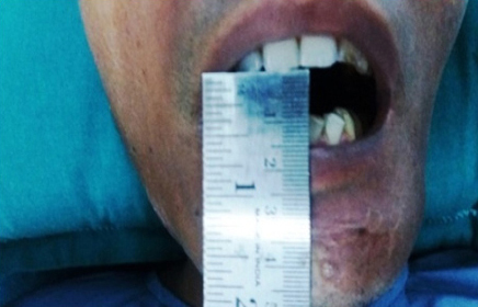
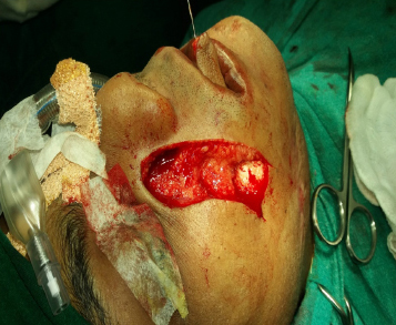
A 25 year old male patient reported to the department of Oral and Maxillofacial Surgery, IDS, Bhubaneswar with the complaint of inability to open mouth properly since the last 3 years. He had a habit of chewing areca nut and catechu for the last 7 years. He did not discontinue his habit until advised to do so and on examination, had a mouth opening of maximal interincisal distance of 10mm (Figure 1). On intraoral palpation there were severe fibrotic bands bilaterally both horizontally and vertically in the buccal mucosa though no ulceration or any exophytic growth was present intraorally.
|
Figure 1: Preoperative interincisal distance measuring about10-15 Mm
Click here to view |
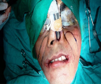
The patient was graded as stage 4 A according to Andrade classification and planned for surgical release of fibrotic bands and inferiorly based melolabial flap reconstruction bilaterally under general anaesthesia.
The patient was nasoendotrachealy intubated by fiberoptic video intubation method due to the extensive limited mouth opening and surgical excision of the fibrotic bands was achieved by combination of scalpel and electrocautery. Upon release of the bands the mouth opening achieved was 36 mm thus obviating the need for coronoidectomy.
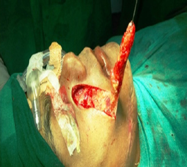
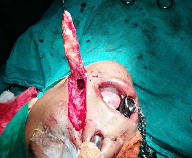
Bilateral inferiorly based melolabial flap marking was done (Figure 2), flap was harvested (Figure 3 and 4) and transbuccal transfer of the flap was done through a tunnel created over the excised defect in the buccal mucosa which covered both the anterior and posterior defects (Figure 5). Tissue excised was sent for histopathological examination to rule out any malignant changes.
|
Figure 2: Markings of bilateral melolabial flap
Click here to view |
|
Figure 3: Raising the flap on right side
Click here to view |
|
Figure 4: Raising the flap on left side
Click here to view |
|
Figure 5: Transbuccal transfer of flap
Click here to view |
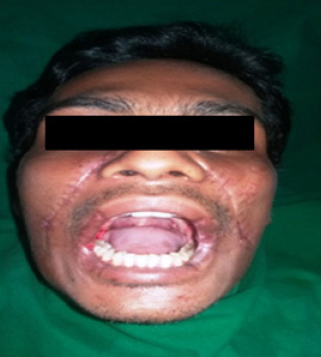
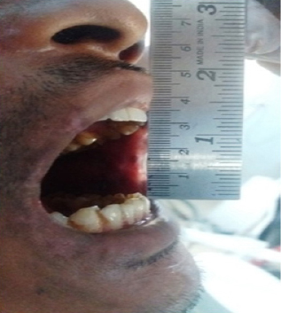
Both the flaps were secured to the defect with 4.0 resorbable sutures (Figure 6) and a gauze pack bung was secured to prevent any untoward complications and was removed on 4th post operative day. The patient was on prophylactic antibiotic therapy and nasogastric feeding for the post operative period. The melolabial flap donor site was meticulously closed using 4.0 prolene non absorbable sutures which was removed on 5th post operative day. Progressive oral physiotherapy using icecream sticks was started on 5th post operative day and the maxmal interincisal distance measured was 30 mm on 14th post operative day (Figure 7 and 8). Mucosalization of the melolabial flap was also seen and patient was informed for the need for eplication in future and scar revision in the region of melolabial flap donor site.

|
Figure 6: Securing the flap to the defect
Click here to view |
|
Figure 7: Post operative picture showing improved mouth opening after 1 week
Click here to view |
|
Figure 8: Post operative picture showing increased interincisal distance.
Click here to view |
The histopathological specimen revealed no malignant transformation though features of OSMF were persistant. The patient was recalled on a progressive schedule to note any complications and was also monitored for the maximum interincisal opening.
DISCUSSION
The areca nut is considered the main factor causing OSMF. The areca nut generates free radicals and causes local immunosuppression along with decreased collagenolysis capability of the mucosa.9 Other etiology and causative agents have also being in literature.10, 11
Melolabial flap is a random pattern flap nurtured on the vascular supply from the transverse facial and angular vessels for the inferior and superiorly based flap respectively. It is a simple, effective, and safe flap. Donor site morbidity is very rare. It effectively covers both posterior and anterior defect of fibrotomy in cases of OSMF and due to its mucosalization it rarely caused post operative contracture and refibrosis.
Surgical descriptions about melolabial flap were found as early as 1830 when Dieffenbach used melolabial flaps to reconstruct nasal alar defects. In 1864, Von Langenbeck used the melolabial flap to reconstruct the nose following ablative therapy. Inferiorly based meolabial flap is a reliable, economical option for the management of oral submucous fibrosis.
The most common complication with the use of melolabial flap is the presence of extraoral scar and intraoral hair growth over the flap. Garatia et al recommended the use of a cheek rotation flap for aesthetic postoperative scar revision.12
Various literature have supported buccal fat pad as a reconstructive option for post fibrotomy reconstruction due to its easy harvesting nature and esthetic purpose. Anshul Rai compared the effectiveness of both melolabial flap and buccal fat pad in a series of fibrotomy cases and concluded the use of buccal fat pad due to its aesthetic efficacy.13 Pradhan et al did a comparative study to evaluate buccal fat pad and collagen sheets for defect closure and concluded collagen sheet is superior in outcome compared to buccal fat pad.14
Borle R on his 25 advanced OSMF case series did melolabial flap reconstruction along with coronoidectomy and concluded it being an effective reconstructive option though the concern being the presence of extra oral scars.15
CONCLUSION
The inferiorly based melolabial flap is a good alternative option for reconstruction of OSMF fibrotomy defects. The external melolabial scar is not devastating and when properly corrected as it lies hidden in the melolabial crease though it can be revised surgically at a later stage.