
INTRODUCTION
John Langdon Down first described DS condition in 1887 and is known as Trisomy 21.1 It is a genetic disorder caused by the presence of all or part of a third copy of chromosome numbered 2.2 The incidence of DS is reported to be 1 in 600 to 1 in 700 live births. During early pregnancy the affected fetuses may even abort.3 Mental retardation will be seen in almost every individual with this syndrome. Many other abnormal clinical traits can also exist along with the characteristic orodental features, like Alzheimer’s disease, leukemia, congenital heart disease, laryngeal atresia. These traits may differ in their severity from individual to individual.4 This syndrome can be identified in a newborn by direct observation or in fetus by prenatal scanning.
CASE REPORT
A 5-year-old male patient, reported to the Department of Oral Medicine & Radiology with the chief complaint of decayed teeth in the upper as well as the lower teeth regions. The child was diagnosed with DS by his physician with no systemic abnormalities and was not on any medication. On eliciting further history, the couple had a consanguineous marriage. The child was born when the mother was 33 years old and was of first order birth. His personal history revealed that he brushes once daily with a toothbrush and toothpaste in a circular motion supervised by his mother. Patient had delayed milestones of growth and immunization schedule was completed. Patient attended school with normal kids and was not able to read or write well at school as revealed by his parents. Mental retardation was present as the patient was nonresponsive to any verbally asked questions and was irritated.
On general examination, patient was moderately built and nourished; height and weight appeared to be normal with his chronological age. Nails, sclera, conjunctiva appeared to be normal. Skin appeared to be dry and rough. Temperomanndibular joint, salivary glands, paranasal sinuses, lymph nodes appeared to be normal. There were no significant systemic problems till date. There was simian crease seen on the dorsal surface of both the palms and the feet appeared to be small and short.
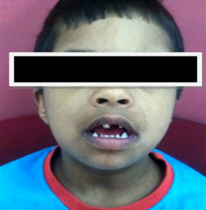
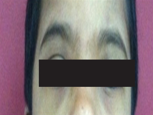
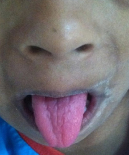
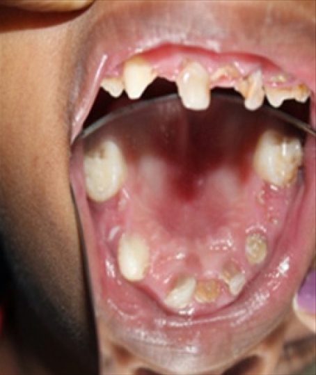
Extra oral examination demonstrated a prominent, broad forehead, a brachycephalic skull, saddle nose deformity, retruded maxilla and protruded mandible. Lips were incompetent, appeared rough and cracked as the patient was a mouth breather. Patient had an open mouth posture with slurred speech (Figure 1). Eyes were almond shaped, ocular hypotelorism, and strabismus was present (Figure 2). Intraoral examination revealed macroglossia with fissures on the dorsum of the tongue, high arch and V shaped palate (Figure 3 and 4). Bleeding on probing was present and rolled out margins of the gingiva was observed. There was generalized spacing with clinically missing 52, root stumps in relation to 54, 61 and dental caries in 53 and 55 along with deep dental caries and associated tenderness with 54 and 85. Conical shaped tooth irt 73 was also seen. Hypodontia and microdontia was present, occlusion showed mesial step terminal plane with an anterior open bite. There was mild calculus and stains present on the teeth.
|
Figure 1: Clinical picture showing an open mouth posture.
Click here to view |
|
Figure 2: Clinical picture revealing strabismus and ocular hypotelorism
Click here to view |
|
Figure 3: Intra oral examination revealing fissured tongue.
Click here to view |
|
Figure 4: Intra oral examination showing a high arch palate
Click here to view |
Based on the history and above described clinical features, a provisional diagnosis of Down’s syndrome was made. A differential diagnosis of Patau (13 chromosome Trisomy) and Edward syndrome (18 chromosome Trisomy) was considered.
As the patient was non cooperative for any treatment planned, basic radiographic investigations were scheduled for the following visits. The patient was then referred to the department of Pedodontics and Preventive dentistry for the further dental treatment. The patient was graded as ’Negative behavior’ given as per Frankel behavior rating scale.4 A step wise scheduled treatment protocol was done for the child.
Initially non-invasive treatment was performed followed by invasive, in order to accustom the patient to the dental environment.
Non-invasive procedures performed were:
Fluoride varnish for all teeth
Pit and fissure sealants for all deciduous molars
Glass ionomer restorations irt 55, 53
Pulpectomy irt 84, 85
Extraction of 61, 63 and space maintainers were advised.
However all the invasive procedures were planned to be done either under conscious sedation or general anesthesia subsequently only after obtaining a physician’s consent. The patient is under a close follow up.
DISCUSSION
Most cases of trisomy 21 (94%) are caused by nondisjunction, resulting in an extra chromosome. The translocation type occurs in 3%, mosaicism occurs in 2%, and rare chromosome aberrations make up the remaining 1% of cases. The incidence of DS is highly correlated with the maternal age; the condition has higher chances of occurrence with increasing maternal age as seen with the present case.3 The oral health of these patients is greatly affected by their medical and physiologic characteristics, thus affecting their quality of life directly or indirectly.5
DS patients have very typical clinical features and most of them were seen in the present case. Orodental Features:
Individuals with DS present with about “35% to 55% of microdontia in both the primary and secondary dentition. Patient’s clinical crowns appear morphologically different than the normal dentition. Malalignment of teeth can also be seen either in permanent or deciduous type of dentition.1 60-63% of DS individuals may have one or more missing teeth.6 An open-mouth posture poses many problems; incompetent lips, difficulties with speech and swallowing.1 All these features were seen in the present case. These patients may also have macroglossia. Tongue appears to be large and protuberant due to reduced oral space in these patients. Fissures may be present on the dorsum of the tongue as seen in the present case.6 Maxillary arch is usually underdeveloped and mandibular arches may be either normal or hypoplastic. They may have a high arched, typical V-shaped palate.6
DENTAL MANAGEMENT
Children with DS should be motivated on a regular oral hygiene regimen. Frequent dietary counseling must be given to prevent any further problems.6 Pit and fissure sealants, topical fluoride application must be done periodically. Periodic oral prophylaxis and periodontal treatment must be done to maintain oral hygiene and an antiseptic mouthwash can be advised.
Dentist must be clear with his communication and give appropriate instructions to them.6 Importance must be given for the maintenance phase rather than the surgical phase. Care must be taken by the pediatrician and pedodontist during any anesthesia procedures. Dental treatment under general anaesthesia is difficult to perform, due to a large protuberant tongue, high arched palate, small mouth, short, broad neck, abnormal dentition, small maxilla & mandible, large tonsils.7
The present case is a unique case report in which usually patients with DS are cooperative but not with the present case. They accept the treatment readily unlike the present case. Due to his negative behavior, many appointments were given and treatment planning was done accordingly.
CONCLUSION
DS patients are a special group of individuals who are affected with severe systemic and oral health problems. They have a mild to a severe degree of retardation and present a challenge to medical practioners and dentists than individuals with any other handicapping conditions. However due to poor resources in many countries, it is becomes difficult to treat DS patients and maintain their oral and overall health in a good condition. By providing good accessibility and improving the treatment facilities the quality of life will become better in these individuals. A dentist can definitely play a significant role in managing these special children effectively and efficiently by following simple treatment procedures.