
INTRODUCTION
Amelogenesis Imperfecta (AI) is a multifarious assemblage of inherited conditions that disturbs the developing enamel structure and presents independent of any related systemic disorders.1 AI is also known as hereditary enamel dysplasia, hereditary brown enamel, and hereditary brown opalescent teeth.2 AI had been first reported in 1890. It was categorized to be distinct clinical entity from dentinogenesis imperfect by Finn (1938).3 Prevalence is highly varied ranging from one in 718 to one in 14,000 depending on the population studied. No racial predilection has been reported.4 The enamel in both primary and permanent dentition is affected. The pattern of inheritance is usually autosomal dominant or recessive, or X linked or sporadic in nature.5
Various classifications have evolved based on phenotype and clinical findings and radiographic appearance. The most widely used classification of AI includes four major categories based primarily on phenotype (hypoplastic [HP], hypomaturation [HM], hypocalcified [HC], HMHP with taurodontism) and subdivided into 15 subtypes based on clinical phenotype and mode of inheritance.6 Clinical presentation of enamel varies from yellowish white discoloration, snow capped enamel, mottled appearance, pits or vertical ridges, hypo mineralized enamel causing it to be abraded from the teeth shortly after their emergence into the oral cavity. AI patients may experience compromised chewing function due to tooth sensitivity and the short clinical crowns caused by attrition and/or incomplete eruption. In the affected teeth, the dentin and root form are normal with low caries susceptibility. 7
Treating a patient with AI is important not only from a functional standpoint, but also from a psychological point of view and hence the aim of the treatment should be to both restore aesthetics, improve masticatory function and to eliminate the adverse social impact. This paper elaborates rehabilitation of a 14 year old girl child diagnosed with hypoplastic form of AI by a minimally invasive approach.
CASE REPORT
A 14-year-old female patient reported to the Department of Pedodontics and Preventive Dentistry, Kamineni Institute of Dental Sciences, Narketpally with the chief complaint of yellowish stains on all teeth which were associated with sensitivity on intake of hot and cold foods. Patient had these discoloured teeth since childhood, and her milk teeth were similarly discoloured too. Patient reported that she felt extreme dissatisfaction with the un-aesthetic appearance of her teeth. The child s parents emphasized on her incapacitating smile and deprived social interaction. This was the patients first dental visit. Past medical and personal history was non-contributory. Positive family history was given where her father and sister had a similar developmental condition. No para-functional habits were reported. Her oral hygiene habits involved brushing once daily using tooth paste and tooth brush in horizontal method.
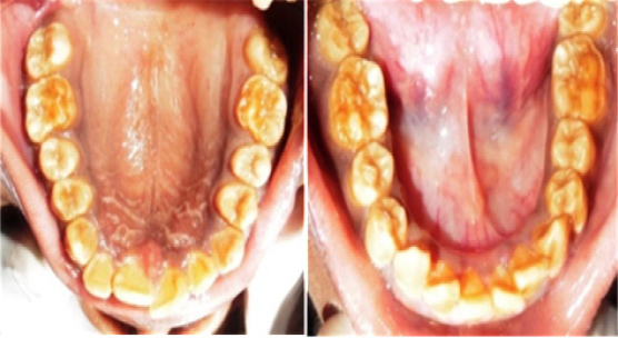
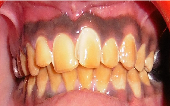
Extra-oral examination revealed straight profile with competent lips. On intra-oral examination, there was full set of permanent dentition with generalized yellowish discoloration and attrition of molars resulting in reduced vertical dimension (Figure 1). Class I malocclusion with anterior deep bite was observed. The emergence pattern and timing of teeth were normal. The oral hygiene status of the patient was fair and examination of soft tissue revealed chronic generalized gingivitis. On probing tooth material was soft in consistency with mild flaking of residual enamel.
|
Figure 1: Intraoral view showing full set of permanent dentition with generalized yellowish discoloration and attrition of molars.
Click here to view |
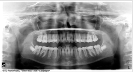
A provisional diagnosis of AI-Hypoplastic type was made based on the history and after clinical examination. Radiographic investigations included an oral pantomograph (OPG) depicted a generalized loss of enamel thickness relating to molars. Radio density of enamel was same as dentin with normal pulp chamber and root morphology. The presence of all unerupted third molars was also seen (Figure 2). Based on history, clinical findings, radiographic examination AI hypo plastic type was given as the diagnosis. As the patient is in her growing stage, treatment is usually carried out in two phases, temporary phase followed by transitory phase. The complexity of the condition requires an interdisciplinary approach for optimal treatment outcome. The treatment objectives were prioritized to conserve the tooth structure, increase the lost vertical dimension to a comfortable position (physiologic neuromuscular position), to restore masticatory function and improve aesthetics. Hence, the treatment was planned in three phases.
|
Figure 2: OPG showing radio-density of enamel was same as dentin with normal pulp chamber and root morphology
Click here to view |
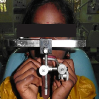
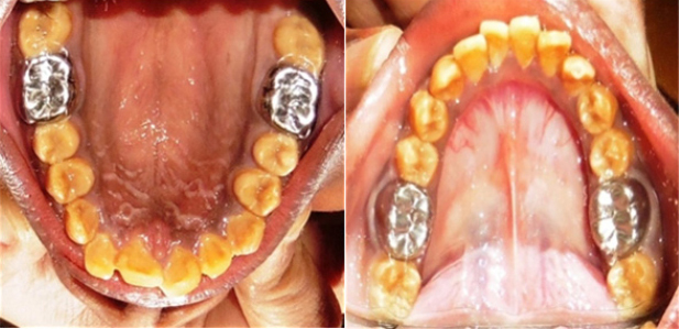
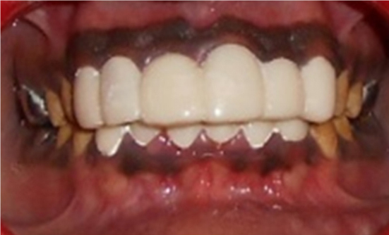
In the first phase the patient was placed on intensive oral hygiene program, including scaling, and after 2 weeks the level of oral hygiene maintained by the patient was acceptable with improvement in soft tissues. Face bow transfer was made to record the cranio-maxillary and maxilla - mandibular relationship and it was then mounted on a semi adjustable articulator which implicated an increase of vertical height by 2mm (Figure 3). In the second phase stainless steel crowns were prepared using minimal preparation making only proximal clearance and cemented with Type I GIC on all four permanent first molars (Figure 4). This treatment provided the necessary full coverage and protected them from further attrition and wear. It also provided an added advantage of increase in patient s vertical dimension by 1mm of occlusion facilitating easy rehabilitation of anterior teeth. Considering age of the patient and partial eruption status of 2nd permanent molars further correction of deep bite was planned at later phase of treatment. On recall after 4 weeks the patient did not have any discomfort so it was decided to move on to the next phase of treatment where the upper and lower anterior teeth were prepared to receive the provisional restorations in the form of heat cure acrylic resin veneers considering the age of the patient (Figure 5 & 6). A definitive treatment requiring placement of porcelain veneers for anterior teeth and metal ceramic crowns for posterior to facilitate full mouth rehabilitation is planned at a later stage.
|
Figure 3: Face-bow transfer.
Click here to view |
|
Figure 4: Stainless Steel Crowns in relation to 16, 26, 36 and 46.
Click here to view |
|
Figure 5: Preparation for heat cure acrylic laminates
Click here to view |
|
Figure 6: Post-operative picture showing heat cure acrylic laminates.
Click here to view |
DISCUSSION
Amelogenesis imperfecta (AI) is a term for a clinically and genetically heterogeneous group of conditions that affect the dental enamel, occasionally in conjunction with other dental, oral and extraoral tissues.8 The most widely accepted classification system for AI is the one described by Witkop in 1989 which divides AI into 3 basic types: hypoplastic;
hypomaturation; and hypocalcified.6 This classification considers the clinical aspects and inheritance pattern and comprises 3 major groups, each with a large number of subtypes. There may be cases in which clinical features of different types of AI coexist. Thus, classifying a patient as having one specific type of AI is a major challenge to the clinician. Some authors have reported that genetic mappings can be a useful tool in identifying to which AI group a patient belongs.9-12 First, one has to exclude the presence of systemic diseases that may show generalized enamel hypoplasia. Secondly, it enables affected families to seek genetic counseling. Finally, an accurate diagnosis helps in recognition of the condition so preventive measures can be provided at an early stage.13 In the present case the patient was diagnosed to be of AI hypoplastic type based on generalized enamel hypoplasia of the permanent teeth with no association of any systemic illness but a positive family history.
In Amelogenesis imperfect with generalized white to yellowish brown discoloration of teeth poses problems of socialization, function, and discomfort. Patients have been known to cover their teeth with pieces of paper, chewing gum or other materials in order to mimic an ordinary” appearance. Adolescents in particular tend to become reclusive and withdrawn, because of their disfigured teeth. The treatment plan should accommodate factors including patient s age, socioeconomic status, disease type, severity, and overall oral condition.14 Treatment planning must focus on early diagnosis, pain management, prevention and restoration of enamel defects, and long-term management. Poor oral hygiene is a recognized problem in patients with AI, because of the rough enamel surface which causes plaque retention and to the sensitivity experienced when brushing. Attention should first be given to the patient s level of oral hygiene and dietary habits, which can compromise the rehabilitation procedures.15 Therefore, oral prophylaxis and oral hygiene instructions, were instituted in the first phase of the presented treatment.
Historically, patients with severe AI have been treated with multiple extractions of the primary teeth followed by the construction of complete dentures.16 Nowadays, there is a range of materials used to restore the teeth that includes composite resin, polycarbonate crowns, stainless steel crowns (SSCs), glass-ionomer cement and functional maintenance dispositives to restore a mutilated dentition.15 In most cases, full coverage is desirable for posterior teeth due to the extensive loss of enamel and also to prevent further loss of tooth structure17. In the present case because of the patient s deep bite and the desire to provide full coverage to all the teeth, Porcelain fused to metal (PFM) crowns were discussed as an option to provide both full coverage and strength; however, the economic impact of treating all the teeth, with the possibility of having to retreat them in the future, made this treatment alternative less desirable.
Stainless Steel Crowns (SSCs) are the most effective type of restoration for posterior teeth as they are extremely durable, relatively cheap, subject to minimal technique sensitivity during placement, and offers the advantage of full coronal coverage.15, 17 In the present case SSCs were used to treat all four permanent second molars. This treatment provided the necessary full coverage and also aided correction of patient s deep bite which made it easier to restore the anterior segments. Scott Rosenblum used Stainless steel crowns in an amelogenesis patient with deep bite.18 The Stainless steel crowns placed on the permanent first molars also facilitated to halt attrition of the occlusal surfaces and decrease the chance of loss of vertical dimension in the future, as well as to protect the dentinal- pulp complex from chemical and thermal attacks.18 Similar technique of using SSCs for rehabilitation of permanent molars in patients with erupting molars was practised by several authors. As this takes development of the child s teeth, their pulpal morphology and the skeletal jaw growth into consideration.151719
In order to restore esthetics of anterior teeth the treatment proposed ranged from bonded veneers restorations to tooth preparations for placement of a full crown. For many years, the most predictable and durable aesthetic restoration of anterior teeth has been achieved with complete crowns. However, this approach requires the preparation of a substantial amount of tooth structure In AI, presence of thin enamel indicates a very conservative preparation and difference of etched enamel pattern compromising the durability of direct composite restoration as indicated by Seow laminates were planned for the patient. Since introduction for a definitive treatment the porcelain laminate veneers gained popularity, as the tooth preparation is conservative and the restorations are aesthetic.20 For the present case, heat cure acrylic veneers were placed to recover esthetics while using a conservative approach which at a later stage could be replaced by porcelain veneers.
The entire treatment duration was approximately 3 months. Systemic and sequential interdisciplinary approach was followed to attain both functional and aesthetic goals to the satisfaction of the patient. This boosted the patient s self-confidence and also increased his responsibility to maintain adequate levels of oral hygiene.
CONCLUSION
Paediatric dentists are the first to encounter children with AI and it is imperative that treatment requires an overall comprehensive plan that includes a rough draft of future treatment needs. Since they are not conditions caused by neglect and cannot, yet, be prevented, it behoves the dental profession to exercise their best skills to alleviate the physco- social conflicts.